In this collaboration, Abberior GmbH and Evotec (Pospiech et al.) combined super-resolution STED microscopy with spatial transcriptomics to decode podocyte injury in human kidney biopsies from Evotec’s Molecular Patient Database (E.MPD).1 STED imaging together with spatial transcriptomics resolved slit diaphragm architecture at nanoscale resolution and linked podocyte effacement to underlying spatial gene expression patterns.
This case study revealed molecular signatures of podocyte effacement and uncovered disease-driving mechanisms - demonstrating how high-resolution imaging and multi-omics generate mechanistic insight and support target discovery in kidney disease.
Decoding Podocyte Effacement: A High-Resolution Approach Using STED Microscopy and Spatial Transcriptomics
Podocytopathies are a heterogeneous group of proteinuric kidney diseases characterized by primary injury to podocytes, the specialized epithelial cells critical for maintaining the integrity of the glomerular filtration barrier.2 These injuries may arise from various sources, including genetic mutations, autoimmune mechanisms, infections, or exposure to toxins. 2 Early morphological changes such as podocyte foot process effacement and disruption of the slit diaphragm precede the onset of proteinuria and represent shared pathogenic mechanisms across various glomerular disorders.2 The histopathological spectrum of podocytopathies ranges from these subtle, early changes undetectable by conventional light microscopy to overt lesions such as segmental sclerosis.3 This variability contributes to diagnostic uncertainty and often necessitates complementary analyses using electron microscopy or immunohistochemistry.3
Recent advances in super-resolution light microscopy, including stimulated emission depletion (STED) and three-dimensional structured illumination microscopy (3D-SIM), enable nanoscale visualization of podocyte foot processes previously achievable only by electron microscopy.4 These techniques not only facilitate more accurate and rapid diagnosis from routine biopsy material but also allow quantitative assessment of slit diaphragm integrity and foot process morphology, offering new insights into the cellular mechanisms underlying podocyte effacement.4
To advance the mechanistic understanding of podocyte injury and effacement, we have collaborated with Abberior GmbH, a leader in STED technology, to combine super-resolution fluorescence microscopy with high-resolution spatial transcriptomics. This integrative study enables parallel visualization of slit diaphragm architecture and spatial gene expression in human kidney biopsies from the NURTuRE patient cohorts.5,6 By linking structural alterations in the glomerular compartment to spatial gene expression signatures, we aim to uncover disease-driving mechanisms and identify novel therapeutic targets to advance precision medicine in glomerular diseases.
Podocytes and other glomerular cell types are often underrepresented in kidney transcriptomic datasets, where signals from proximal tubules and other abundant tubulointerstitial cell populations dominate.7 To specifically investigate the molecular mechanisms driving glomerular disease progression, we performed high-resolution spatial transcriptomics (Visium HD, 10x Genomics) on formalin-fixed, paraffin-embedded kidney biopsies from healthy living donors (n = 3), patients with idiopathic nephrotic syndrome (INS, n = 8), and chronic kidney disease (CKD, n = 8). Hematoxylin and eosin (H&E) staining enabled expert histopathological classification of 186 glomeruli into normal morphology, mesangial expansion, glomerulosclerosis, or other etiology-specific phenotypes (Figure 1A).
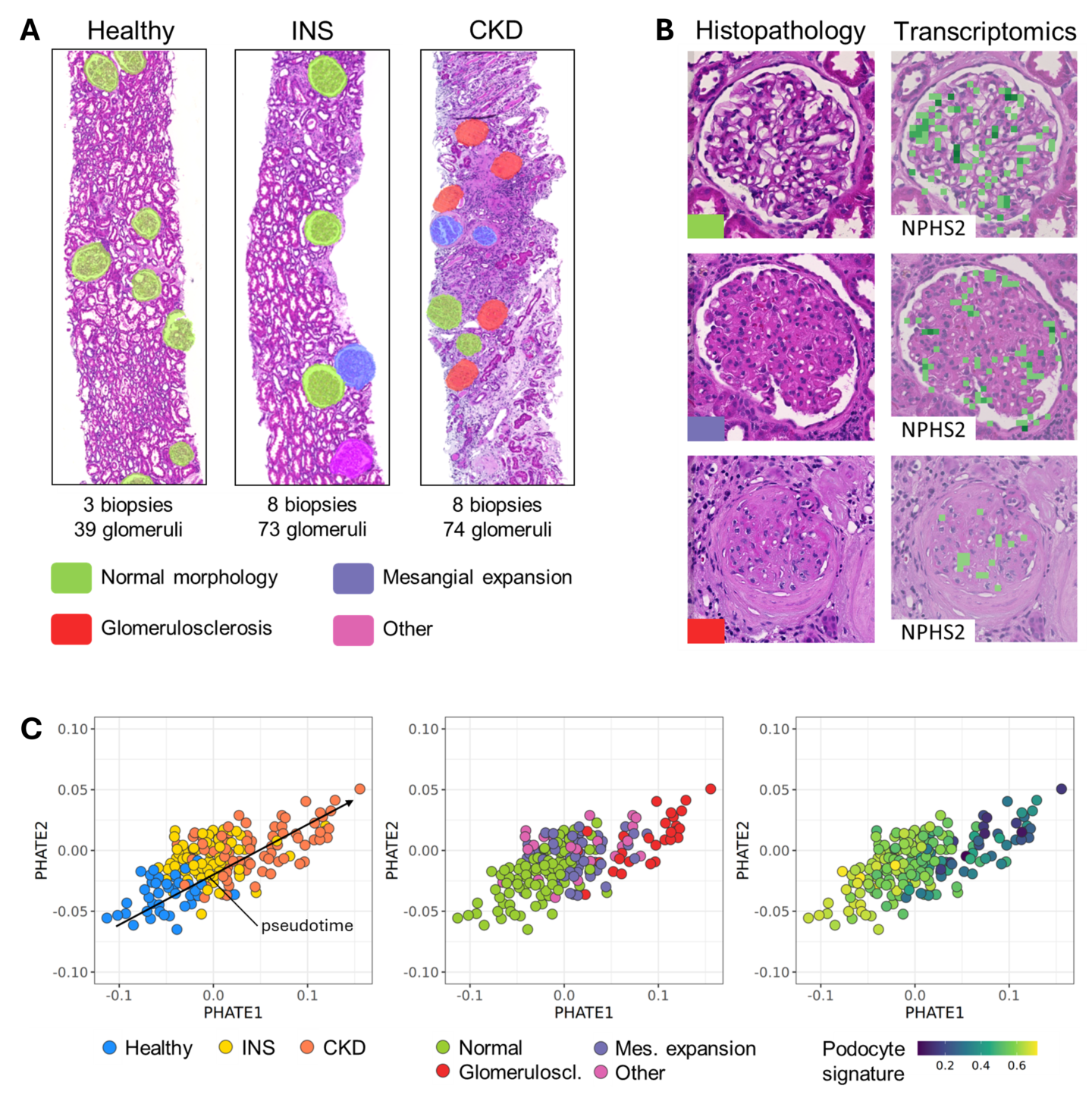
Figure 1: High-resolution spatial transcriptomics and expert histopathology analysis of patient kidney biopsies revealed a molecular trajectory of glomerular disease progression. (A) Kidney biopsies were obtained from healthy living kidney donors (healthy, n = 3), patients with idiopathic nephrotic syndrome (INS, n = 8), and chronic kidney disease (CKD, n = 8) and subjected to hematoxylin and eosin (HE) staining and high-resolution spatial transcriptomics analysis (Visium HD, 10x Genomics) on consecutive formalin-fixed and paraffin-embedded tissue sections. A kidney histopathology expert classified a total of 186 glomeruli (39 healthy, 73 INS, and 74 CKD) into normal morphology (green), mesangial expansion (purple), glomerulosclerosis (red), or other (pink) disease-specific phenotypes. (B) Representative HE images and spatial transcriptomics data documenting the loss of podocyte marker expression (podocin, NPHS2) in glomeruli with mesangial expansion and glomerulosclerosis. Spatial gene expression data were binned into 8 x 8 µm squares and overlaid with the HE images. (C) PHATE dimension reduction of pseudobulk transcriptomes representing a molecular trajectory of glomerular disease progression. Spatial gene expression data from 186 glomerular entities were aggregated into individual pseudobulk transcriptomes. Annotation of glomerular pseudobulk transcriptomes with clinical and histopathological features aligned with the molecular trajectory capturing pseudotime gene expression changes associated with mesangial expansion, glomerulosclerosis and loss of podocytes.
Spatial transcriptomics revealed a progressive loss of the podocyte marker podocin (NPHS2) in glomeruli exhibiting mesangial expansion and glomerulosclerosis (Figure 1B). To capture global transcriptional changes associated with glomerular remodeling, we aggregated spatial gene expression data from individual glomeruli into pseudobulk transcriptomes and applied PHATE dimension reduction.8 This analysis uncovered a continuous molecular trajectory of glomerular disease progression that aligned with histopathological classifications and clinical annotations. The trajectory captured pseudotime-associated transcriptional changes linked to mesangial matrix expansion, glomerulosclerosis, and podocyte depletion, providing critical context for analyzing podocyte effacement and slit diaphragm integrity across different glomerular disease stages (Figure 1C).
To investigate structural and molecular correlates of podocyte injury along this trajectory, we performed super-resolution STED microscopy on consecutive sections for a subset of 80 glomeruli from the spatial transcriptomics cohort. This subset was enriched for glomeruli with normal H&E morphology, enabling focused analysis of early structural alterations associated with primary podocyte injury. STED imaging of nephrin (NPHS1) revealed distinct slit diaphragm morphologies, which were classified into normal, diffuse global, or segmental effacement patterns (Figure 2A). Diffuse global effacement was frequently observed in glomeruli with normal H&E morphology and associated with INS, consistent with early, primary podocyte injury. In contrast, segmental effacement was more commonly observed in glomeruli with mesangial expansion or glomerulosclerosis, suggesting secondary podocyte injury in the context of CKD.
To resolve the molecular landscape of podocyte effacement at higher spatial resolution, we applied the BANKSY niche segmentation algorithm to identify intra-glomerular tissue domains enriched for podocytes.9 PHATE dimension reduction of these podocyte niche transcriptomes revealed a clear separation between niches with normal slit diaphragm morphology and those with diffuse global effacement (Figure 2B). Differential gene expression analysis identified 49 upregulated and 341 downregulated transcripts in globally effaced niches, with enrichment for pathways involved in slit diaphragm maintenance, including cell junction organization, actin cytoskeleton dynamics, and endocytosis. Consistent with the structural disruption observed by STED microscopy, expression of nephrin (NPHS1), a key component of the slit diaphragm, was markedly reduced in effaced niches, while podocalyxin (PODXL) expression was upregulated, potentially reflecting altered podocyte polarity or compensatory changes in surface composition.
Our integrative approach combining super-resolution STED microscopy with high-resolution spatial transcriptomics revealed a molecular trajectory that captures the continuum of glomerular disease progression, including mechanisms of podocyte depletion, mesangial expansion, and glomerulosclerosis. By resolving structural and transcriptional changes at unprecedented spatial resolution, we identified a unique molecular signature of podocyte foot process effacement and uncovered disease-driving pathways within the glomerular compartment. This study demonstrates how collaboration and cutting-edge technology can drive scientific innovation in kidney research, providing a foundation for mechanistic disease understanding and the identification of new precision therapeutics in glomerular diseases.
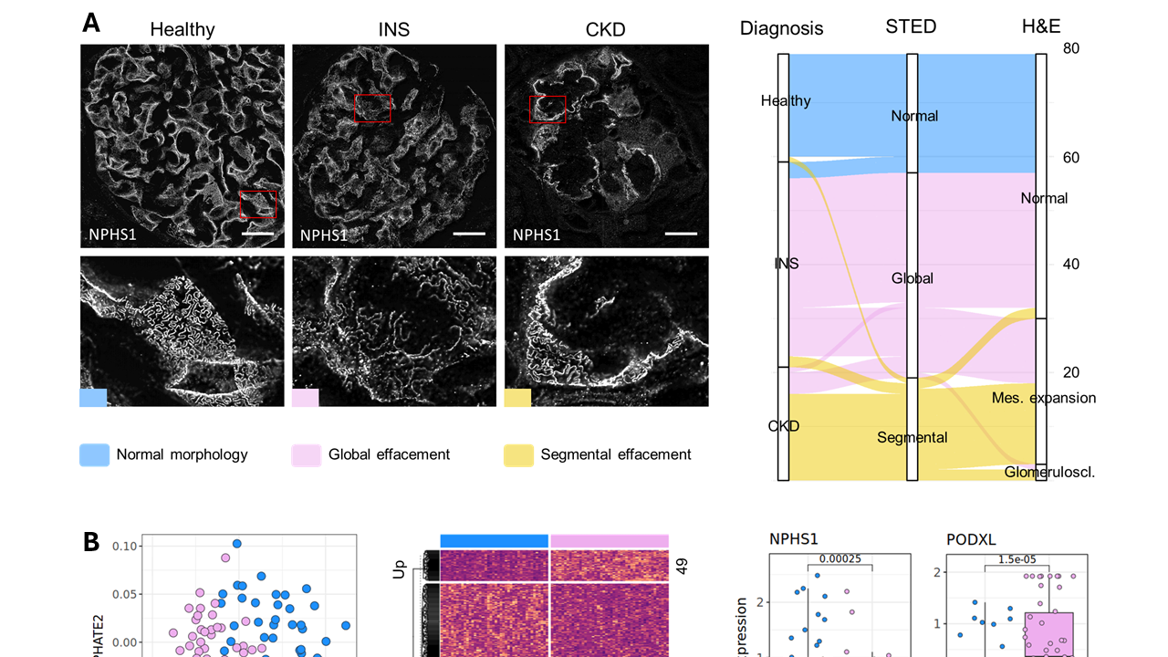
Figure 2: High-resolution spatial transcriptomics and stimulated emission depletion (STED) microscopy analysis of patient kidney biopsies revealed a unique molecular signature associated with podocyte foot process effacement. (A) Representative stimulated emission depletion (STED) microscopy images of the glomerular filtration slit in kidney biopsies from healthy living donors (healthy), idiopathic nephrotic syndrome (INS), and chronic kidney disease (CKD) patients. The glomerular filtration slit was visualized by nephrin (NPHS1) immunostaining and STED microscopy and subjected to expert histopathology assessment. Glomeruli were classified by appearance of their filtration slits into normal morphology (blue), diffuse global (pink) or segmental (yellow) podocyte effacement. The filtration slit STED and H&E morphology were summarized by diagnosis groups in an alluvial plot, highlighting the association of diffuse global podocyte effacement with normal H&E morphology and INS. (B) PHATE dimension reduction of podocyte niche transcriptomes representing the molecular disease landscape separating niches with normal morphology (blue) from niches with diffuse global foot process effacement (pink). Differential gene expression analysis between normal and globally effaced podocyte niches resulting in 49 upregulated and 341 downregulated transcripts was summarized in a heatmap. Differentially expressed genes were enriched for mechanisms associated with the maintenance of the slit diaphragm, including regulation of cell junctions, actin cytoskeleton and endocytosis (not shown). Nephrin (NPHS1) and podocalyxin (PODXL) expression are shown as examples.
References
- Pospiech, J. et al. Decoding podocyte effacement: A high-resolution approach using STED microscopy and spatial transcriptomics. Nephrology Dialysis Transplantation 40, gfaf116.0359 (2025).
- Kopp, J. B. et al. Podocytopathies. Nat Rev Dis Primers 6, 68 (2020).
- Ravaglia, F. et al. The Pathology Lesion Patterns of Podocytopathies: How and why? Front Cell Dev Biol 10, 838272 (2022).
- Siegerist, F., Campbell, K. N. & Endlich, N. A new era in nephrology: the role of super-resolution microscopy in research, medical diagnostic, and drug discovery. Kidney Int 107, 994–1001 (2025).
- Taal, M. W. et al. Associations with age and glomerular filtration rate in a referred population with chronic kidney disease: methods and baseline data from a UK multicentre cohort study (NURTuRE-CKD). Nephrology Dialysis Transplantation 38, 2617–2626 (2023).
- Colby, E. et al. National Unified Renal Translational Research Enterprise: Idiopathic Nephrotic Syndrome (NURTuRE-INS) study. Clin Kidney J 17, sfae096 (2024).
- Lake, B. B. et al. An atlas of healthy and injured cell states and niches in the human kidney. Nature 619, 585–594 (2023).
- Moon, K. R. et al. Visualizing structure and transitions in high-dimensional biological data. Nat Biotechnol 37, 1482–1492 (2019).
- Singhal, V. et al. BANKSY unifies cell typing and tissue domain segmentation for scalable spatial omics data analysis. Nat Genet 56, 431–441 (2024).