Introduction
- The environment created by a 3D culture model allows reconstitution of the natural cellular physiology by promoting the complex cell-cell and cell-matrix network communications found in vivo.
- Different scaffold-free 3D microtissue models are available at Cyprotex including spheroids produced using ultra-low adhesion plates.
- Microtissues can be formed from single or co-cultured cell populations and their longevity permits use in long-term repeat dose toxicity studies to investigate any cumulative effect.
- ATP content is a biochemical endpoint used to measure general cell viability. This can be incorporated with LDH release to monitor cell death over time.
Protocol
ATP Content in 3D Microtissues
Data
Data from ATP Content in 3D Microtissues Assay
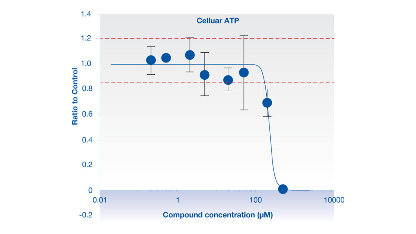
Figure 1
Determination of cellular ATP levels using Promega’s CellTiter Glo® luminescent cell viability assay, following exposure of 3D microtissues to rosiglitazone.