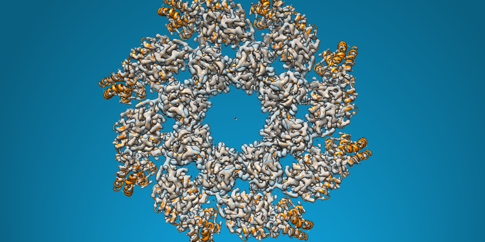
Cryo-electron microscopy (cryo-EM) is a leading technique for high-resolution structure determination of complex biological assemblies, particularly membrane proteins and protein–nucleic acid complexes. By capturing proteins in near-native states without crystallization, cryo-EM has transformed structural biology. It is ideal for targets that are difficult to analyze using conventional methods, revealing dynamic conformations and heterogeneous samples. As a result, cryo-EM plays a vital role in modern drug discovery, offering deep structural insights that support rational design and molecular understanding.
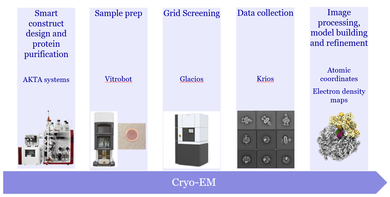
Cryo-EM workflow at Evotec – from sample prep to structural insight
A Complete Solution For Cryo-EM Projects
- Construct and complex formation design focuses on stabilizing proteins, increasing particle size, and reducing preferential orientation during sample preparation. This supports high-resolution structure determination with cryo-EM, using Fabs and nanobodies when needed
- Negative stain analysis identifies protein size, shape, oligomeric state, and aggregation, providing essential early insights before cryo-EM imaging
- Cryo-EM screening and data acquisition follow rapid, optimized workflows to produce high-quality vitrified grids. Evotec ensures access to advanced cryo-EM microscopes and detectors for consistent imaging
- Image processing and model refinement are customized to each project, using specialized software to deliver detailed structural data and validation
Specialised Membrane Protein Solutions
- Broad expertise across challenging membrane targets, including GPCRs, ion channels, solute carriers, and bacterial proteins
- Use of rigid, IP-free tagging strategies and detergent/lipid screening to stabilize smaller proteins and GPCR systems for monodisperse, thermostable preparations
- Tailored optimization strategies to improve particle distribution on cryo-EM grids and enhance resolution of membrane domains during data processing